Responsible: Dr. Antoni Mestre
Ictal SPECT
The ictal perfusion SPECT is performed after the reduction or suspension of antiepileptic drugs in patients admitted to the Epilepsy Monitoring Unit. The tracer administration, 18-30 mCi (666-1,110 MBq) of 99mTc HMPAO or 99mTcECD is performed intravenously in the first 20 seconds of the onset of a seizure under VEEG monitoring. The acquisition of the images is carried out after the stabilization of the patient in a period between 15 and 90 minutes after the end of the seizure. The images are acquired in a Siemens ECAM double-head collimator with high resolution, 128 × 128 matrix, acquisition of 120 projections of 30 seconds. The images are reconstructed by rear projection filtered using a Butterworth filter.
Interictal SPECT
Interictal perfusion SPECT was performed under pharmacological treatment, with the same procedure as in ictal SPECT, but in the absence of ictal activity during the tracer injection.
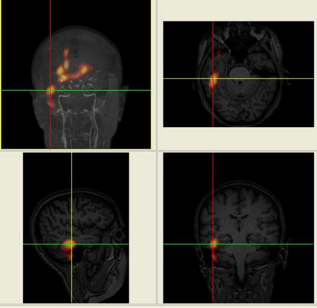
SISCOM
This is a post-processing of the images using the methodology Substraction of Ictal SPECT Co-register to MRI (SISCOM). SISCOM combines the functional information of the SPECT with the anatomical information of the MRI, performs the subtraction of the ictal and interictal SPECT and the subsequent co-registration in the MRI. Currently two possible processing programs are used: Analyze 7.0 (Biomedical Imaging Resource, Mayo Foundation, Rochester, MN, USA) and FocusDET (www.ciber-bbn.es/focusdet).
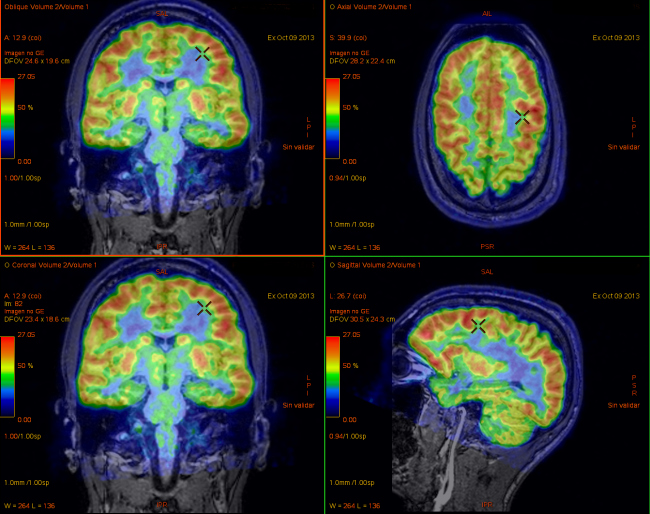
PET 18F-FDG
The PET18F-FDG or FDG PET is performed in an interictal situation and evaluates the cerebral metabolism at baseline (absence of seizure) under pharmacological treatment. It is performed by a 4-crown PET / CT scanner ST scanner (General Electric Medical Systems) at 40-45 minutes after intravenous administration of a dose of 5 MBq / kg of 18 F-FDG. The acquisition of the images is done in three-dimensional (3D) mode with attenuation correction by means of a TC 120-130 Kv acquisition. The reconstruction of the images is done according to the iterative algorithm Ordered Subset Expectation Maximization (OSEM).
Co-registration or PET-RM Fusion
Image fusion of FDG PET with MRI is currently performed using a processing program that allows the integration of images from both studies using a mathematical model (AW Server 2.0). A joint visual assessment of the nuclear physician and neuroradiologist is performed.
References:
- Perfusion SPECT, SISCOM and PET (18)F-FDG in the assessment of drug- refractory epilepsy patients candidates for epilepsy surgery. Suárez-Piñera M, Mestre-Fusco A, Ley M, González S, Medrano S, Principe A, Mojal S, Conesa G, Rocamora R. Rev Esp Med Nucl Imagen Mol. 2015 Nov-Dec;34(6):350-7.
- Garibotto V, Picard F. Nuclear medicine imaging in epilepsy. Epileptologie.2013;30:109-21.
- Goffin K, Dedeurwaerdere S, van Laere K, van Paesschen W. Neuronuclear assessment of patients with epilepsy. Semin Nucl Med. 2008;38:227-39.13.
- Clinical Role of Subtraction Ictal SPECT Coregistered to MR Imaging and (18)F-FDG PET in Pediatric Epilepsy. Perissinotti A, Setoain X, Aparicio J, Rubí S, Fuster BM, Donaire A, Carreño M, Bargalló N, Rumiá J, Garcia-Fructuoso G, Mayoral M, Sanmartí F, Pons F. J Nucl Med. 2014 Jul;55(7):1099-105.
- Rubí S1, Setoain X, Donaire A, Bargalló N, Sanmartí F, Carreño M, Rumià J, Calvo A, Aparicio J, Campistol J, Pons F. Validation of FDG-PET/MRI coregistration in nonlesional refractory childhood epilepsy. Epilepsia. 2011 Dec;52(12):2216-24.