14 April 2021 - Press notes
Awareness of this fact may provide anaesthesiologists with increased control over the procedure, allow them to carry it out more safely and allow for more accurate dosage of anaesthetics. The study, which has been published by the journal Sleep, was carried out with healthy volunteers who underwent neuroimaging tests to assess the brain's reaction during the anaesthetic procedure. Researchers have been able to determine the exact moment in which the cerebral cortex and brain stem go out of sync, namely, when loss of consciousness is achieved.
Researchers from the Hospital del Mar have been able to determine the exact moment in which patients undergoing anaesthesia lose consciousness. They have been able to detect the sequence through which the brain leaves a state of consciousness and falls into unconsciousness through the use of neuroimaging tests. This breakthrough will allow for more careful monitoring of patients and improved control of anaesthetic procedures.
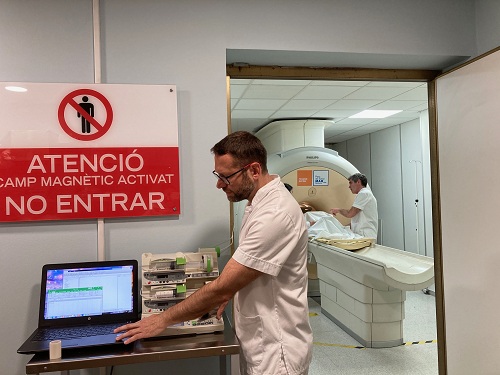
Dr. Juan L. Fernández Candil and Dr. Lluís Gallart
The research published by the journal Sleep and carried out between 2017 and 2018, stems from the collaboration between researchers from the Hospital del Mar's anaesthesiology department, led by Dr. Juan L. Fernández Candil, and a group from the Magnetic Resonance Unit from the same hospital's radiology department, led by Dr. Jesús Pujol. The test was carried out on 21 healthy volunteers, who were anaesthetised with propofol. While the drug was being administered, the volunteers were required to press a sensor every two seconds to track their loss of consciousness. Meanwhile, their vital signs were monitored and their brain activity was tracked through magnetic resonance and an encephalogram.
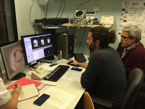
Gerard Martínez-Vilavella and Dr. Jesús Pujol
Brain images obtained through magnetic resonance showed how there was a loss of connection between the cerebral cortex, which is in charge of the brain's executive functions, and the subcortical part and the brain stem, at the moment in which volunteers ceased to press their hands, that is, when they lost consciousness. This is the first time that this precise moment has been identified with images and records.
Dr. Juan L. Fernández Candil, an attending physician from the anaesthesiology department and one of the authors of the paper, points out that "up until now we had devices that helped us identify the approximate moment when the patient is unconscious, but not the exact moment in which they lose consciousness." In some cases, this could lead to an excess dose of anaesthetics to ensure that patients remain in a state of unconsciousness, with all the ensuing problems that entails.
However, the results open the way for more accurate information regarding patients' threshold of consciousness, thus improving safety and control over the procedure and ensuring that patients do not retain any memories of the procedures they may undergo. As Dr. Luís Gallart, the head of the anaesthesiology department and another co-author of the work points out, "calibrating the patient's situation has always been tricky, and there is a risk of overdosage. If you have a monitor that allows you to adjust anaesthetic doses to know if patients are conscious or unconscious, this could help limit overdosage, and therefore the possible side effects of anaesthetics." The side effects in question are mainly delirium and post-operative cognitive decline.
Researchers will now continue to analyse the data they have obtained in order to validate a method to provide their discoveries with practical applications in the operating room by monitoring patients' states of consciousness through the information observed in their encephalograms. Dr. Fernández Candil states that "we cannot have a magnetic resonance in every operating room, but we could have a device that allows us to monitor a patient's encephalogram. Thus, by correlating the results from our clinical tests (when patients stopped pressing with their hands) and neuroimaging with the encephalograms of the volunteers who participated in our research, we would obtain a valid tool to know when patients lose consciousness". Moreover, "this would allow us to administer much more accurate doses, improve the safety of the procedure and provide more guarantees that the patient is completely unconscious throughout surgery."
Pujol J, Blanco-Hinojo L, Gallart L, Moltó L, Martínez-Vilavella G, Vilà E, Pacreu S, Adalid I, Deus J, Pérez-Sola V, Fernández-Candil J. Largest scale dissociation of brain activity at propofol-induced loss of consciousness. Sleep. 2021 Jan 21;44(1):zsaa152. doi: 10.1093/sleep/zsaa152. PMID: 32813022.


 This site complies with the HONcode standard for trustworthy health information:
 Parc Salut Mar
Passeig Marítim 25-29 Barcelona 08003
See location on Google maps
Phone: 93 248 30 00 · Fax: 93 248 32 54
Information request
© 2006 - 2024 Parc de Salut Mar · Legal notice and Privacy Police | Cookie Policy | Accessibility