The SEEG is an invasive diagnostic technique developed in France in the 1960s by Bancaud and Talairach in the Sainte-Anne hospital. It involves the implantation of multiple microelectrodes using stereotactic technique. Designing a correct implantation scheme requires many years of practice and experience. Its goal is the evaluation of an anatomo-electro-clinical correlate in the generation and propagation of seizures. Therefore, the analysis of these records allows us to know the spatio-temporal dynamics of epileptic seizures related to semiology with a high degree of anatomical precision.
SEEG Indications
-
Polymicrogyria (PMG)
-
Tuberous Sclerosis (TSC)
-
Focal Cortical Dysplasia (CFD)
-
Periventricular Heterotopias (HPV)
-
RNM negative (not lesional)
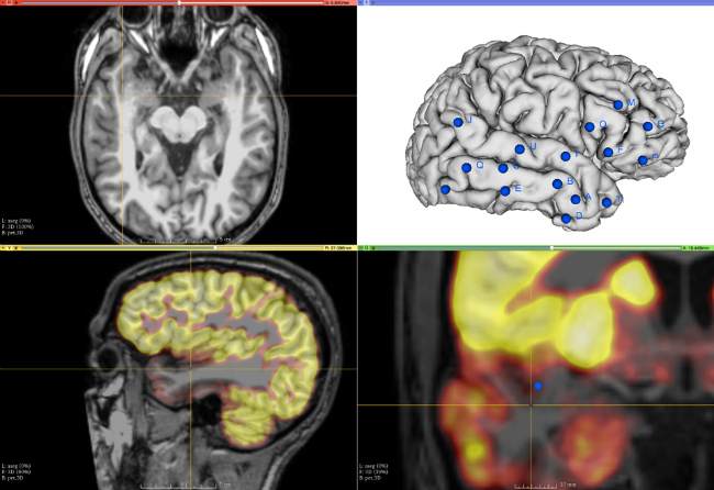
Implementation planning using 3DSlicer and multimodal analysis PET-RM.

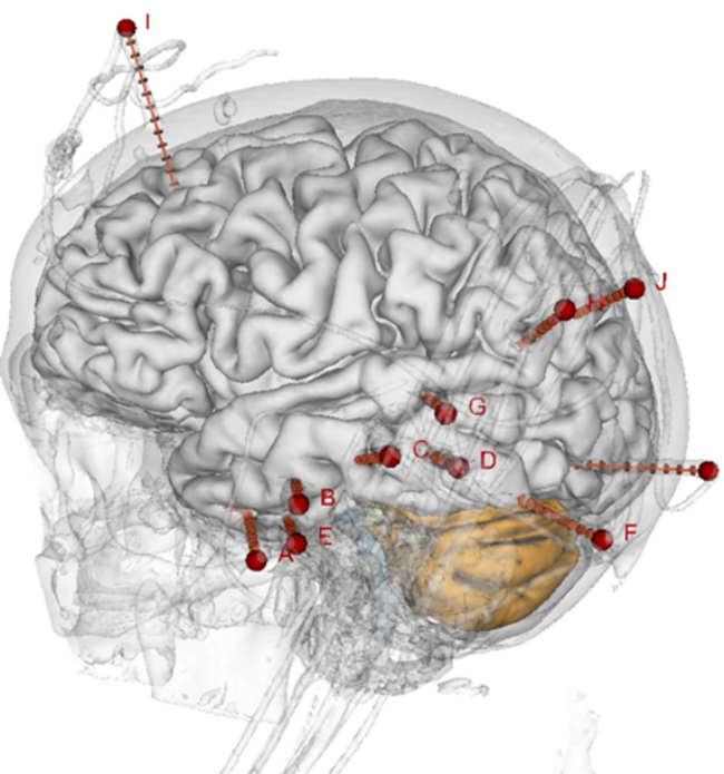
SEEG Postprocessing for accurate electrode location using Freesurfer and MNI atlases.
Example of SEEG in non-lesional temporal epilepsy.
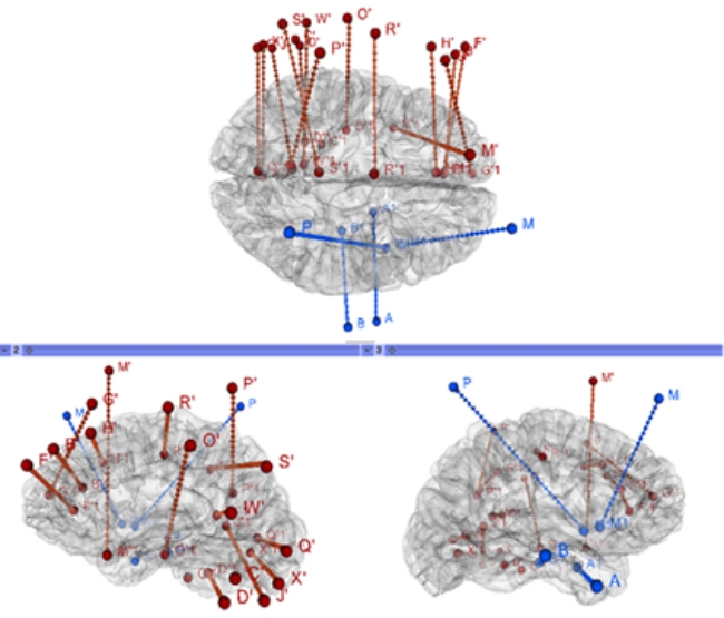
Example of bilateral SEEG in insular epilepsy.